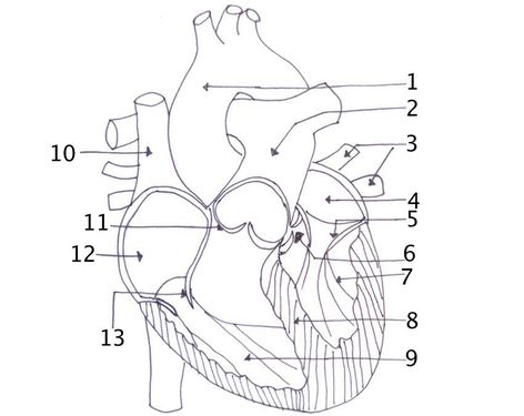
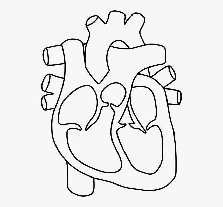
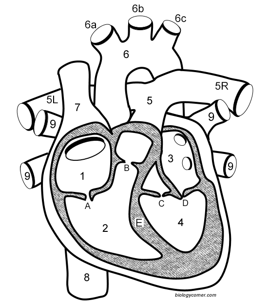
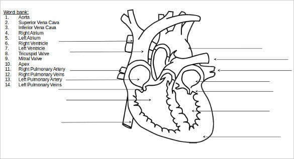
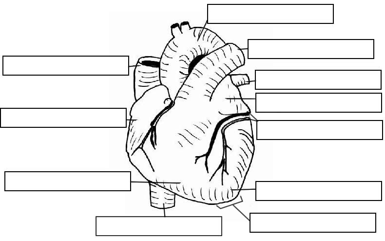
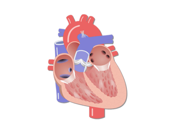
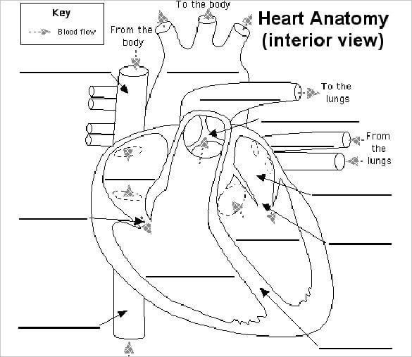
38 structure of the heart without labels
ebook - Wikipedia An ebook (short for electronic book), also known as an e-book or eBook, is a book publication made available in digital form, consisting of text, images, or both, readable on the flat-panel display of computers or other electronic devices. WebAIM: WebAIM's WCAG 2 Checklist Feb 26, 2021 · 2.4.6 Headings and Labels (Level AA) Page headings and labels for form and interactive controls are informative. Avoid duplicating heading (e.g., "More Details") or label text (e.g., "First Name") unless the structure provides adequate differentiation between them. 2.4.7 Focus Visible (Level AA)
Structure of the Heart | The Franklin Institute The heart consists of four chambers: two atria on the top and two ventricles on the bottom. Looking at the Valentine's Day heart, the two rounded humps at the top are rounded like the top of a lower-case "a." The bottom is shaped like a "v." Feel it working What else is inside your heart?
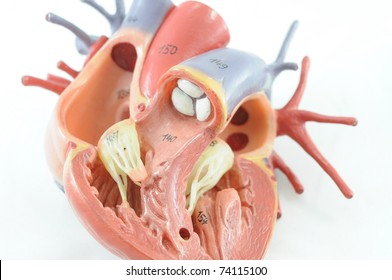
Structure of the heart without labels
the heart without labels Label the heart - Teaching resources. 17 Pics about Label the heart - Teaching resources : 35 Label The Parts Of The Heart - Labels For You, Heart without labels and also 35 Label The Parts Of The Heart - Labels For You. Label The Heart - Teaching Resources wordwall.net label heart diagram labelled Quia - Label The Heart Heart Chambers Without Labels / Label Every Structure On The Figure Of ... The 4 valves are the aortic, . The basic anatomy and physiology of the heart. Heart anatomy · right and left sides of the heart · superior and inferior vena cavae · pulmonary arteries · pulmonary veins · aorta · valves · coronary . If you sever all the nerves to the heart, . In this interactive, you can label parts of the human heart. Human Ear Diagram - Bodytomy The Structure of Human Ear. Helix: It is the prominent outer rim of the external ear. Antihelix: It is the cartilage curve that is situated parallel to the helix. Crus of the Helix: It is the landmark of the outer ear, situated right above the pointy protrusion known as the tragus. Auditory Ossicles: The three small bones in the middle ear ...
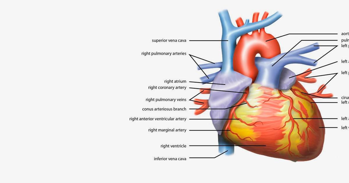
Structure of the heart without labels. Human Heart Diagram Labeled | Science Trends The heart's atrioventricular valves are structures that join the atria and ventricles of the heart together. This group of valves is comprised of the tricuspid valve and the mitral valve. Beyond this, there is a structure referred to as the aortic valve which separates the left ventricle and the aorta. Heart Anatomy Labeling Game - PurposeGames.com This is an online quiz called Heart Anatomy Labeling Game There is a printable worksheet available for download here so you can take the quiz with pen and paper. Your Skills & Rank Total Points 0 Get started! Today's Rank -- 0 Today 's Points One of us! Game Points 19 You need to get 100% to score the 19 points available Actions Structure and Function of the Heart - News-Medical.net Structure of the heart. The heart wall is composed of three layers, including the outer epicardium (thin layer), middle myocardium (thick layer), and innermost endocardium (thin layer). The ... Human Heart (Anatomy): Diagram, Function, Chambers, Location in Body The heart is a muscular organ about the size of a fist, located just behind and slightly left of the breastbone. The heart pumps blood through the network of arteries and veins called the...
Heart without labels - SlideShare 1 of 9 Heart without labels Apr. 08, 2011 • 7 likes • 7,878 views Download Now Download to read offline Education anatomy of heart from bsn class, but now modified without the labels so you can practice. John Martin Follow Lab Assistant Advertisement Recommended 150 Heart RONALDO QUITCO Ap2 chap18heartclass MissReith Anatomy of the heart TLS - Times Literary Supplement Our critics review new novels, stories and translations from around the world Diagram of Human Heart and Blood Circulation in It Four Chambers of the Heart and Blood Circulation The shape of the human heart is like an upside-down pear, weighing between 7-15 ounces, and is little larger than the size of the fist. It is located between the lungs, in the middle of the chest, behind and slightly to the left of the breast bone. The Anatomy of the Heart, Its Structures, and Functions - ThoughtCo The heart is the organ that helps supply blood and oxygen to all parts of the body. It is divided by a partition (or septum) into two halves. The halves are, in turn, divided into four chambers. The heart is situated within the chest cavity and surrounded by a fluid-filled sac called the pericardium. This amazing muscle produces electrical ...
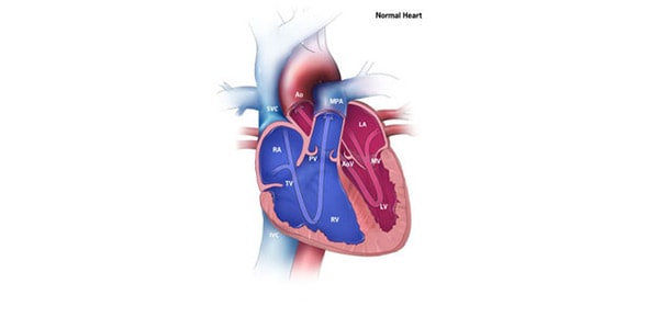
Human Heart Diagram Without Labels - Labelling Worksheet - Twinkl The human heart is a muscle made up of four chambers, these are: Two upper chambers - the left atrium and right atrium Two lower chambers - the left and right ventricles. It's also made up of four valves - these are known as the tricuspid, pulmonary, mitral and aortic valves. Structure of the Heart | Biology for Majors II - Lumen Learning In humans, the heart is about the size of a clenched fist, and it is divided into four chambers: two atria and two ventricles. There is one atrium and one ventricle on the right side and one atrium and one ventricle on the left side. The atria are the chambers that receive blood, and the ventricles are the chambers that pump blood. Heart Diagram with Labels and Detailed Explanation - BYJUS Diagram of Heart. The human heart is the most crucial organ of the human body. It pumps blood from the heart to different parts of the body and back to the heart. The most common heart attack symptoms or warning signs are chest pain, breathlessness, nausea, sweating etc. The diagram of heart is beneficial for Class 10 and 12 and is frequently ... Heart (Human Anatomy): Overview, Function & Structure | Biology The heart is a muscular organ that pumps blood throughout the body. It is located in the middle cavity of the chest, between the lungs. In most people, the heart is located on the left side of the chest, beneath the breastbone. The heart is composed of smooth muscle. It has four chambers which contract in a specific order, allowing the human ...
Microsoft takes the gloves off as it battles Sony for its ... Oct 12, 2022 · Microsoft pleaded for its deal on the day of the Phase 2 decision last month, but now the gloves are well and truly off. Microsoft describes the CMA’s concerns as “misplaced” and says that ...
Heart: Anatomy and Function - Cleveland Clinic The parts of your heart are like the parts of a house. Your heart has: Walls. Chambers (rooms). Valves (doors). Blood vessels (plumbing). Electrical conduction system (electricity). Heart walls Your heart walls are the muscles that contract (squeeze) and relax to send blood throughout your body.
Human Heart Diagram Without Labels - Pinterest Heart Diagram Learning Objectives 1. Describe the three major layers of the heart wall and how they relate to the pericardium; identify the three layers of arteries and veins. 2. Identify and describe the function of the chambers of the heart, the […] W Laura Gleba heart
DNA - Wikipedia The structure of DNA is dynamic along its length, being capable of coiling into tight loops and other shapes. In all species it is composed of two helical chains, bound to each other by hydrogen bonds. Both chains are coiled around the same axis, and have the same pitch of 34 ångströms (3.4 nm
human heart diagram without labels Label Kidney Diagram - Human Anatomy tartrerepub.blogspot.com. label kidney urinary anatomy diagram vessels system renal human lab models torso corpuscle sdmesa classroom edu. Structure Of The Heart - YouTube . diagram heart easy sketch structure 1364 paintingvalley healthiack. Draw The Core 516.2.1
Label the heart — Science Learning Hub In this interactive, you can label parts of the human heart. Drag and drop the text labels onto the boxes next to the diagram. Selecting or hovering over a box will highlight each area in the diagram. pulmonary vein semilunar valve right ventricle right atrium vena cava left atrium pulmonary artery aorta left ventricle Download Exercise Tweet
Heart anatomy: Structure, valves, coronary vessels | Kenhub Heart anatomy. The heart has five surfaces: base (posterior), diaphragmatic (inferior), sternocostal (anterior), and left and right pulmonary surfaces. It also has several margins: right, left, superior, and inferior: The right margin is the small section of the right atrium that extends between the superior and inferior vena cava .
Structure Of The Heart | A-Level Biology Revision Notes The heart is a hollow muscular organ that lies in the middle of the chest cavity. It is enclosed in the pericardium, which protects the heart and facilitates its pumping action. The heart is divided into four chambers: The two atria (auricles): these are the upper two chambers. They have thin walls which receive blood from veins.
Heart Diagram Unlabeled - Cliparts.co 84 images of Heart Diagram Unlabeled. You can use these free cliparts for your documents, web sites, art projects or presentations. Don't forget to link to this page for attribution!
Internet - Wikipedia It operates without a central governing body. The technical underpinning and standardization of the core protocols (IPv4 and IPv6) is an activity of the Internet Engineering Task Force (IETF), a non-profit organization of loosely affiliated international participants that anyone may associate with by contributing technical expertise.
Sotalol: Uses, Interactions, Mechanism of Action | DrugBank ... Pharmacodynamics. Sotalol is a competitive inhibitor of the rapid potassium channel. 2 This inhibition lengthens the duration of action potentials and the refractory period in the atria and ventricles. 3,4 The inhibition of rapid potassium channels is increases as heart rate decreases, which is why adverse effects like torsades de points is more likely to be seen at lower heart rates. 6 L ...
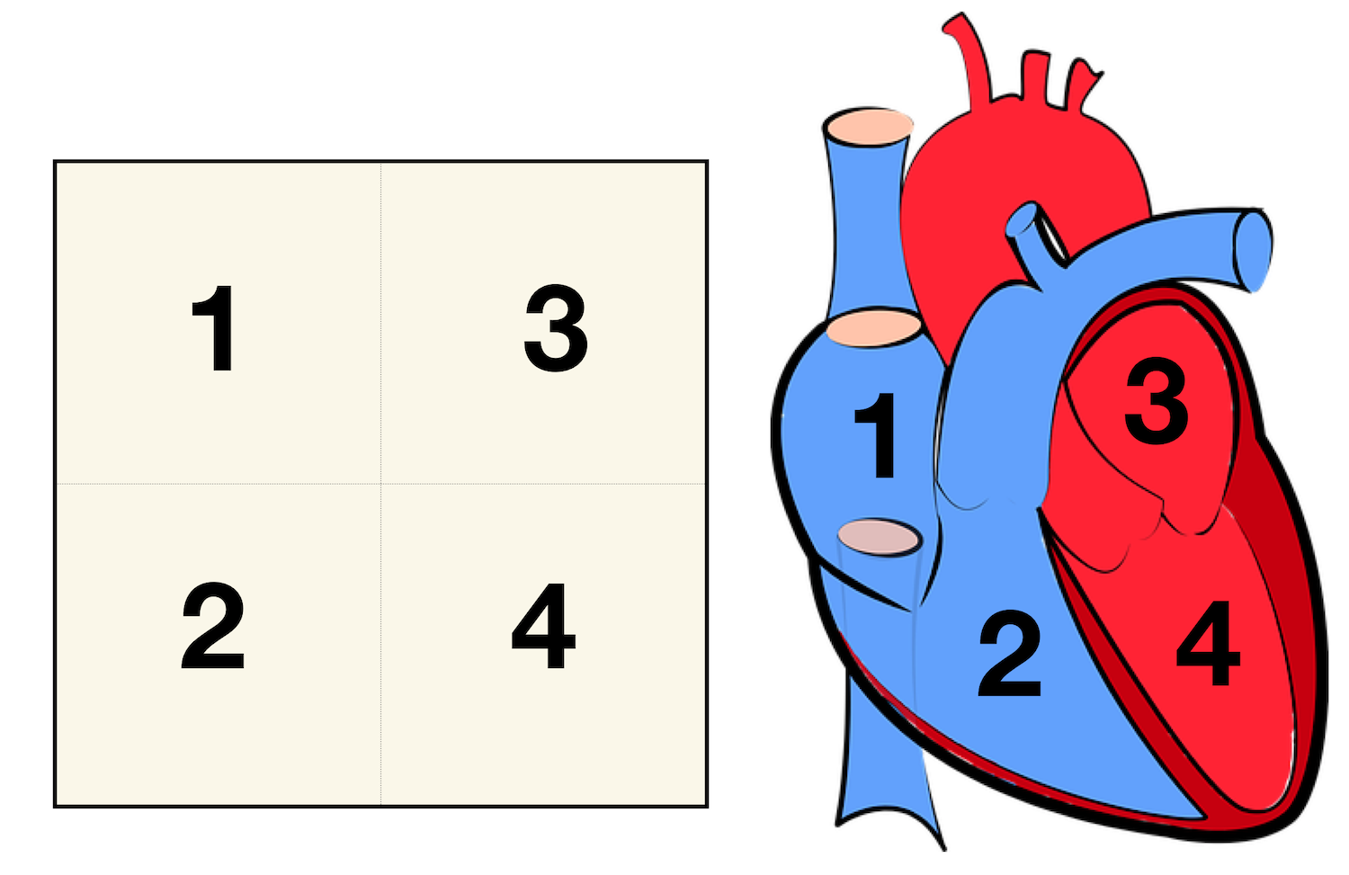
Heart Anatomy: Labeled Diagram, Structures, Blood Flow ... - EZmed Right vs Left Side of the Heart Now that we have converted the heart into a square with 4 different boxes or chambers, the heart can be divided into 2 sides. First, the right side is shown in blue and includes boxes/chambers 1 and 2. The left side is shown in red and includes boxes/chambers 3 and 4. View fullsize
Structure of the Heart | SEER Training - National Cancer Institute The human heart is a four-chambered muscular organ, shaped and sized roughly like a man's closed fist with two-thirds of the mass to the left of midline. The heart is enclosed in a pericardial sac that is lined with the parietal layers of a serous membrane. The visceral layer of the serous membrane forms the epicardium. Layers of the Heart Wall
Anatomy of the heart and blood vessels | Patient Blood vessels form the living system of tubes that carry blood both to and from the heart. All cells in the body need oxygen and the vital nutrients found in blood. Without oxygen and these nutrients, the cells will die. The heart helps to provide oxygen and nutrients to the body's tissues and organs by ensuring a rich supply of blood.
A Labeled Diagram of the Human Heart You Really Need to See The human heart, comprises four chambers: right atrium, left atrium, right ventricle and left ventricle. The two upper chambers are called the left and the right atria, and the two lower chambers are known as the left and the right ventricles. The two atria and ventricles are separated from each other by a muscle wall called 'septum'.
Structure and function of the heart - BBC Bitesize It is located in the middle of the chest and slightly towards the left. The heart is a large muscular pump and is divided into two halves - the right-hand side and the left-hand side. The...
heart | Structure, Function, Diagram, Anatomy, & Facts heart, organ that serves as a pump to circulate the blood. It may be a straight tube, as in spiders and annelid worms, or a somewhat more elaborate structure with one or more receiving chambers (atria) and a main pumping chamber (ventricle), as in mollusks. In fishes the heart is a folded tube, with three or four enlarged areas that correspond to the chambers in the mammalian heart. In animals ...
Human Ear Diagram - Bodytomy The Structure of Human Ear. Helix: It is the prominent outer rim of the external ear. Antihelix: It is the cartilage curve that is situated parallel to the helix. Crus of the Helix: It is the landmark of the outer ear, situated right above the pointy protrusion known as the tragus. Auditory Ossicles: The three small bones in the middle ear ...
Heart Chambers Without Labels / Label Every Structure On The Figure Of ... The 4 valves are the aortic, . The basic anatomy and physiology of the heart. Heart anatomy · right and left sides of the heart · superior and inferior vena cavae · pulmonary arteries · pulmonary veins · aorta · valves · coronary . If you sever all the nerves to the heart, . In this interactive, you can label parts of the human heart.
the heart without labels Label the heart - Teaching resources. 17 Pics about Label the heart - Teaching resources : 35 Label The Parts Of The Heart - Labels For You, Heart without labels and also 35 Label The Parts Of The Heart - Labels For You. Label The Heart - Teaching Resources wordwall.net label heart diagram labelled Quia - Label The Heart


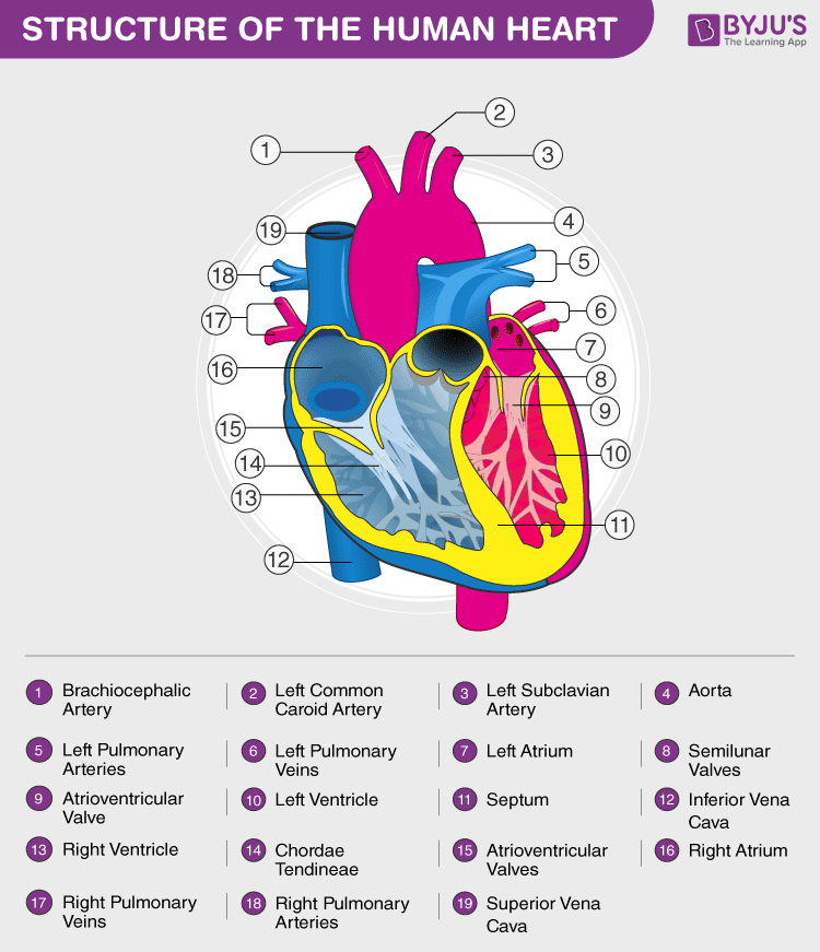

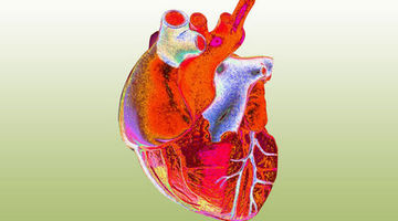
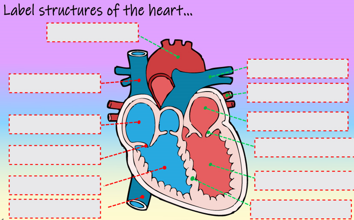












Post a Comment for "38 structure of the heart without labels"