38 external structure of the heart with labels
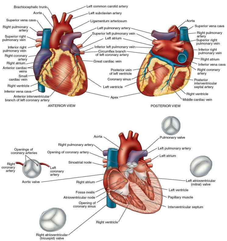
Ch. 19 Circulatory System- heart Flashcards | Quizlet Correctly label the external anatomy of the anterior heart. Place the labels in order denoting the flow of blood through the pulmonary circuit beginning with the right atrium and ending in the left atrioventricular valve. The first and last structures are given. Right atrium 1. tricuspid valve 2. right ventricle 3. pulmonary valve Heart Diagram with Labels and Detailed Explanation - BYJUS Well-Labelled Diagram of Heart. The heart is made up of four chambers: The upper two chambers of the heart are called auricles. The lower two chambers of the heart are called ventricles. The heart wall is made up of three layers: The outer layer of the heart wall is called epicardium. The middle layer of the heart wall is called myocardium.
Heart Labeling Quiz: How Much You Know About Heart Labeling? Here is a Heart labeling quiz for you. The human heart is a vital organ for every human. The more healthy your heart is, the longer the chances you have of surviving, so you better take care of it. Take the following quiz to know how much you know about your heart. Questions and Answers 1. What is #1? 2. What is #2? 3. What is #3? 4. What is #4?

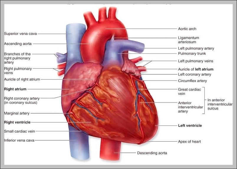
External structure of the heart with labels
en.wikipedia.org › wiki › DapagliflozinDapagliflozin - Wikipedia Medical uses. Dapagliflozin is used along with diet, exercise and usually with other glucose lowering medications, to improve glycaemic control in adults with type 2 diabetes and to reduce the risk of hospitalisation for heart failure among adults with type 2 diabetes and known cardiovascular disease or other cardiovascular risk factors (including high blood pressure, high cholesterol and ... The Anatomy of the Heart, Its Structures, and Functions Cardiac conduction is the rate at which the heart conducts electrical impulses. Heart nodes and nerve fibers play an important role in causing the heart to contract. Atrioventricular Bundle: A bundle of fibers that carry cardiac impulses. Atrioventricular Node: A section of nodal tissue that delays and relays cardiac impulses. › programs › heart-weekHeart Week 2022 | The Heart Foundation Heart Health Checks present a valuable opportunity for healthcare professionals to engage with their patients about their risk of developing cardiovascular disease and ways to lower this risk.
External structure of the heart with labels. quizlet.com › 630625176 › chapter-19-the-heart-flashChapter 19: The Heart Flashcards | Quizlet •Allows heart to beat without friction, gives it room to expand and resists excessive expansion •Parietal pericardium-tough outer, fibrous layer of connective tissue-inner serous layer •Visceral pericardium (a.k.a. epicardium of heart wall)-serous lining of sac turns inward at base of heart to cover the heart surface Heart Anatomy: Heart Dissection - University of Washington External Features of the Heart. The heart is contained within a thin membranous sac, ... The letters indicated in the text refer to the labels on the picture. ... Video 5.1.6 ("Ventricles: outflow pathways") is particularly useful for showing the structure and action of the valves. The action of the valves is shown by pumping water through the ... en.wikipedia.org › wiki › The_TenorsThe Tenors - Wikipedia The Tenors (formerly known as The Canadian Tenors) are a vocal group consisting of Victor Micallef, Fraser Walters, and Clifton Murray.They perform operatic pop music that is a mixture of classical and pop, featuring songs such as "The Prayer", Panis angelicus, and Leonard Cohen's Hallelujah. Heart - External Features - Anatomy QA Three surfaces. Sternocostal surface: Is formed by right atrium, right ventricle, left auricle and left ventricle. Left surface: Is formed by left auricle and left ventricle. Diaphragmatic surface: 2/3rd is formed by left ventricle and 1/3rd by right ventricle. Posterior surface/Base: It is quadrilateral in shape.
Human Heart - Anatomy, Functions and Facts about Heart The heart wall is made up of 3 layers, namely: Epicardium - Epicardium is the outermost layer of the heart. It is composed of a thin-layered membrane that serves to lubricate and protect the outer section. Myocardium - This is a layer of muscle tissue and it constitutes the middle layer wall of the heart. Human Heart - Diagram and Anatomy of the Heart - Innerbody The heart is a muscular organ about the size of a closed fist that functions as the body's circulatory pump. It takes in deoxygenated blood through the veins and delivers it to the lungs for oxygenation before pumping it into the various arteries (which provide oxygen and nutrients to body tissues by transporting the blood throughout the body). Detailed Structure of Frog's Heart - Microbiology Notes Protects the heart from mechanical injury. Keeps the heart moist; Allows the free movement during beating. Also keeps in keeping the heart suspended in its proper position. External Structure of Heart. Externally heart looks like a triangular structure. It is reddish color. It is 3 chambered besides sinus venosus and truncus arteriosus. Development of the Heart | Anatomy and Physiology | | Course Hero From head to tail, strong include the truncus arteriosus, bulbus cordis, primitive ventricle, primitive atrium, and the sinus venosus. Initially, all venous blood flows into the sinus venosus, and contractions propel the blood from tail to head, or from the sinus venosus to the truncus arteriosus.
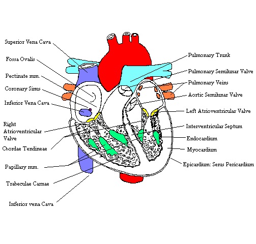
A Labeled Diagram of the Human Heart You Really Need to See The human heart, comprises four chambers: right atrium, left atrium, right ventricle and left ventricle. The two upper chambers are called the left and the right atria, and the two lower chambers are known as the left and the right ventricles. The two atria and ventricles are separated from each other by a muscle wall called 'septum'. The structure of the heart - Structure and function of the heart ... Each side of the heart consists of an atrium and a ventricle which are two connected chambers. The atria (plural of atrium) are where the blood collects when it enters the heart. The ventricles... Heart Anatomy Labeling Game - PurposeGames.com This is an online quiz called Heart Anatomy Labeling Game. There is a printable worksheet available for download here so you can take the quiz with pen and paper. Your Skills & Rank. Total Points. 0. Get started! Today's Rank--0. Today 's Points. One of us! Game Points. 19. You need to get 100% to score the 19 points available. Heart Wall Anatomy | Structure of the Heart Wall | GetBodySmart The epicardium layer of the heart wall. 1 2 The epicardium is a serous membrane that consists of an external layer of simple squamous and an inner layer of areolar tissue ( loose connective tissue ). The squamous cells secrete lubricating fluids into the pericardial cavity. The thick middle layer of the heart wall is called the myocardium.
Structure of the Heart | Heart structure, Medical knowledge, Nursing students
hbr.org › 2009 › 09How Strategy Shapes Structure - Harvard Business Review See Industrial Market Structure and Economic Performance, F. M. Sherer (Chicago: Rand McNally, 1970). 2. See Blue Ocean Strategy , W. Chan Kim and Renée Mauborgne (Harvard Business Press, 2005).

DNA Replication - Structure - Stages of Replication - TeachMePhyiology
Lesson | The Heart - External Structure | Encounter Edu In this lesson students begin their exploration of the circulatory system, labelling a diagram of the external structures and identifying arteries and veins. They will go on to explain where blood enters and leaves the heart. Learning outcomes

Pin on Anatomy Human Other Bambie Reed
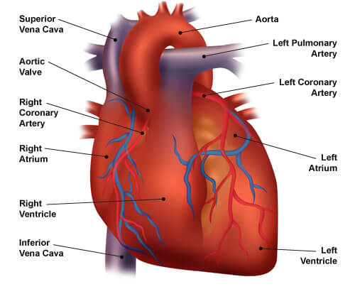
Structure of the Heart Flashcards | Quizlet Structure of the Heart. STUDY. Flashcards. Learn. Write. Spell. Test. PLAY. Match. Gravity. Created by. jay_birdX. Key Concepts: Terms in this set (15) Aorta. The largest artery in the body; it conducts freshly oxygenated blood from the heart to the tissues. ... One of the four heart valves, the tricuspid valve is the first one that blood ...
heart
Heart anatomy: Structure, valves, coronary vessels | Kenhub The heart is shaped as a quadrangular pyramid, and orientated as if the pyramid has fallen onto one of its sides so that its base faces the posterior thoracic wall, and its apex is pointed toward the anterior thoracic wall.

Ongzi's Lifelong Learning: Typhoid Fever
File:Diagram of the human heart (cropped).svg - Wikipedia Add Inferior vena cava and pericardium labels: 18:08, 14 August 2018: 656 × 631 (209 KB) Jmarchn: Add pericardium. Add papillary muscles and chordae tendinae. Add cardiac skeleton. Inferior vena cava more wide. ... Diagram of the human heart, created by Wapcaplet in Sodipodi. Cropped by ~~~ to remove white space (this cropping is not the same ...
.jpg)
Heart Model & Sheep Heart Practice Quiz - ProProfs Quiz
Label the heart — Science Learning Hub Add to collection. In this interactive, you can label parts of the human heart. Drag and drop the text labels onto the boxes next to the diagram. Selecting or hovering over a box will highlight each area in the diagram. Right ventricle. Right atrium. Left atrium. Pulmonary artery. Left ventricle.

The Heart | S-cool, the revision website
› physiology-of-the-heartPhysiology of the Heart | Boundless Anatomy and Physiology ... The first wave on an ECG is the P wave, indicating atrial depolarization in which the atria contract (atrial systole ). The P wave is the first wave on the ECG because the action potential for the heart is generated in the sinoatrial (SA) node, located on the atria, which sends action potentials directly through Bachmann's bundle to depolarize the atrial muscle cells.

The Heart - Biology Student
Structure Of The Heart | A-Level Biology Revision Notes The heart is a hollow muscular organ that lies in the middle of the chest cavity. It is enclosed in the pericardium, which protects the heart and facilitates its pumping action. The heart is divided into four chambers: The two atria (auricles): these are the upper two chambers.

Anatomy Heart Quizlet
Anatomy of a Human Heart - uofmhealth The coronary arteries run along the surface of the heart and provide oxygen-rich blood to the heart muscle. A web of nerve tissue also runs through the heart, conducting the complex signals that govern contraction and relaxation. A sac known as the pericardium surrounds the heart.

Heart anatomy vector illustration - VectorMine
How to Draw the Internal Structure of the Heart (with Pictures) To draw the internal structure of a human heart, follow the steps below. Part 1 Finding a Diagram 1 To find a good diagram, go to Google Images, and type in "The Internal Structure of the Human Heart". Find an image that displays the entire heart, and click on it to enlarge it. 2 Find a piece of paper and something to draw with.
32 Label The Diagram Of The Heart - Label Design Ideas 2020
Layers of the heart: Epicardium, myocardium, endocardium - Kenhub The myocardium is functionally the main constituent of the heart and the thickest layer of all three heart layers. It is a muscle layer that enables heart contractions. Histologically, the myocardium is comprised of cardiomyocytes. Cardiomyocytes have a single nucleus in the center of the cell, which helps to distinguish them from skeletal muscle cells that have multiple nuclei dispersed in the periphery of the cell.
HeartComplete
Structures of the Heart - Biology LibreTexts Figure 40.9. 1: Human Heart: (a) The heart is primarily made of a thick muscle layer, called the myocardium, surrounded by membranes. One-way valves separate the four chambers. (b) Blood vessels of the coronary system, including the coronary arteries and veins, keep the heart muscles oxygenated.
Congestive Heart Failure: The Essence of Heart Failure Course | CEUfast Nursing Continuing Education
draw and label the heart heart draw structure internal human structures external anatomy drawing labeled anatomical wikihow step valves veins left atrium. 34 Label Of Heart Diagram - Labels Database 2020 otrasteel.blogspot.com. heart diagram label system cardiovascular key labels anatomy health human organ. Heart Diagram Drawing At GetDrawings.com | Free For Personal ...

Medical Tourism News and Views: How does the heart work?
moody-challenge.physionet.org › 2022George B. Moody PhysioNet Challenge | Quick links for this ... Feb 22, 2022 · The segmentation annotation file (with .tsv extension and in plain text format) is composed of three distinct columns: the first column corresponds to the time instant (in seconds) where the wave was detected for the first time, the second column corresponds to the time instant (in seconds) where the wave was detected for the last time, and the third column corresponds to an identifier that ...

In this diagram they are showing the function of the heart as they have labels to the ...
Labelling the heart — Science Learning Hub Blood transports oxygen and nutrients to the body. It is also involved in the removal of metabolic wastes. In this interactive, you can label parts of the human heart. Drag and drop the text labels onto the boxes next to the diagram. Selecting or hovering over a box will highlight each area in the diagram.
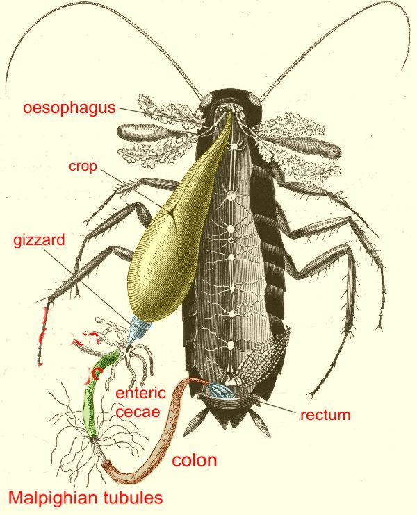
cockroaches parasites 3
Heart - Wikipedia The heart has four chambers, two upper atria, the receiving chambers, and two lower ventricles, the discharging chambers.The atria open into the ventricles via the atrioventricular valves, present in the atrioventricular septum.This distinction is visible also on the surface of the heart as the coronary sulcus. There is an ear-shaped structure in the upper right atrium called the right atrial ...

Labeling the Heart
Structure of the Heart | SEER Training Chambers of the Heart. The internal cavity of the heart is divided into four chambers: Right atrium; Right ventricle; Left atrium; Left ventricle; The two atria are thin-walled chambers that receive blood from the veins. The two ventricles are thick-walled chambers that forcefully pump blood out of the heart. Differences in thickness of the heart chamber walls are due to variations in the amount of myocardium present, which reflects the amount of force each chamber is required to generate.
Post a Comment for "38 external structure of the heart with labels"