43 microscope images with labels
Every IB Biology drawing you NEED to know - YouTube PLEASE READ!This is every single drawing you need to know!Looking through the 2016 syllabus, this video covers 2 main types of statements- Draw- Annotate ( +... Amazon.com: My First Lab Duo Scope Microscope - Young ... KIDS MICROSCOPE KIT -- This kids microscope kit, in addition to the user manual and experiment guide, includes 5 blank slides, 1 concavity (well) slide, 4 prepared slides, 2 bottles of stain, 5 slide labels, 5 cover glasses, 50 sheets of lens paper, 1 plastic transfer pipette, 1 plain wooden applicator, 1 cotton-tipped applicator, 1 plastic ...
Microscope Objective Lens | Products | Leica Microsystems Microscope Objectives. Leica Microsystems – The Ultimate in Optical Competence. For more than 170 years Leica Microsystems has designed and produced top-class objectives for a wide variety of applications in research, industry and medicine. The optics specialists at Leica Microsystems bring the highest level of experience and expertise to bear in reducing …
Microscope images with labels
Electron microscope - Wikipedia An electron microscope is a microscope that uses a beam of accelerated electrons as a source of illumination. As the wavelength of an electron can be up to 100,000 times shorter than that of visible light photons , electron microscopes have a higher resolving power than light microscopes and can reveal the structure of smaller objects. Microscope Slide Label Templates - TemplateMonster Minimalist is a beautiful PowerPoint template you can use to design professional presentations for business and agency meetings and events. The template comes with 140 unique slides in HD resolution and features image placeholders, world maps, icons, and much more.Key Features : 140 different slides, Simple. Parts of Stereo Microscope (Dissecting microscope) - Rs' Science Stereo microscopes (also called Dissecting microscope) are branched out from other light microscopes for the application of viewing "3D" objects. These include substantial specimens, such as insects, feathers, leaves, rocks, sand grains, gems, coins, and stamps, etc. Functionally, a stereo microscope is like a powerful magnifying glass.
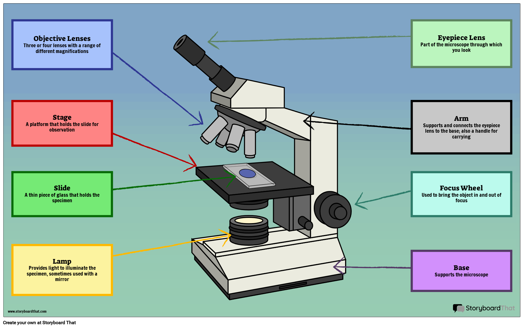
Microscope images with labels. Labeling the Parts of the Microscope Labeling the Parts of the Microscope This activity has been designed for use in homes and schools. Each microscope layout (both blank and the version with answers) are available as PDF downloads. You can view a more in-depth review of each part of the microscope here. Download the Label the Parts of the Microscope PDF printable version here. Smart Microscope for Lab Routine and Research - ZEISS In the past, documenting samples with multiple fluorescent labels in your routine lab could be time consuming. To get best image quality, you needed to manually switch filters, adjust illumination intensities and exposure times and to snap each single channel image. For four different channels, this could sum up to 15 steps and clicks. With Smart Microscopy, this is a … Parts of Stereo Microscope (Dissecting microscope) – labeled … Compared to a compound microscope where the objectives attached to the nosepiece can be seen and identified individually (based on color bands and their respective labels), the objectives of a dissecting microscope are located in a cylindrical cone and, therefore, are not directly seen. For the stereo microscope that comes with multiple objective lens sets (fixed power style), the … Compound Microscope Parts - Labeled Diagram and their Functions - Rs ... The eyepiece (or ocular lens) is the lens part at the top of a microscope that the viewer looks through. The standard eyepiece has a magnification of 10x. You may exchange with an optional eyepiece ranging from 5x - 30x. [In this figure] The structure inside an eyepiece. The current design of the eyepiece is no longer a single convex lens.
26+ Picture Of A Microscope With Label PNG 26+ Picture Of A Microscope With Label PNG. Microscopes are specially created to magnify the image of the subject being studied. Students label the parts of the microscope in this photo of a basic laboratory light microscope. Microscope Drawing And Label at GetDrawings | Free download from getdrawings.com I searched for this on bing.com/images. Electron microscope - Wikipedia An electron microscope is a microscope that uses a beam of accelerated electrons as a source of illumination. As the wavelength of an electron can be up to 100,000 times shorter than that of visible light photons, electron microscopes have a higher resolving power than light microscopes and can reveal the structure of smaller objects.. Electron microscopes use shaped magnetic … Microscope labels | Bill Todt Microscope labels. Unlabelled image: Lab Index: 101 Class Page: Identify the following parts of the microscope: Eyepieces Observation tube Nosepiece Objectives Stand Specimen holder Mechanical stage Stage controls Condenser Iris diaphragm Coarse focus Fine focus Skin Images Labeled | Virtual Anatomy Lab VAL - ncccval Skin Images Labeled | Virtual Anatomy Lab VAL ... Connective Tissue Images Unlabeled. Microscope. Microscope Images Labeled. Microscope Images Unlabeled. Mitosis. Mitosis Images Labeled. Mitosis Images Unlabeled. Skin. Skin Images Labeled. Skin Images Unlabeled. Skeletal system. Skeletal Images Labeled. Skeletal Images Unlabeled.
Microscope Drawing And Label - Painting Valley Are you looking for the best images of Microscope Drawing And Label? Here you are! We collected 33+ Microscope Drawing And Label paintings in our online museum of paintings - PaintingValley.com. ADVERTISEMENT LIMITED OFFER: Get 10 free Shutterstock images - PICK10FREE label microscope diagram compound parts light labeling functions microscopic Microscope Labeled Pictures, Images and Stock Photos Browse 48 microscope labeled stock photos and images available, or start a new search to explore more stock photos and images. Newest results Fluorescent Imaging immunofluorescence of cancer cells growing... Plant Tissue Systems vector illustration. Labeled biology... Microscope diagram vector illustration. Labeled zoom instrument... Parts of a microscope with functions and labeled diagram Optical parts of a microscope and their functions The optical parts of the microscope are used to view, magnify, and produce an image from a specimen placed on a slide. These parts include: Eyepiece - also known as the ocular. This is the part used to look through the microscope. Its found at the top of the microscope. Compound Microscope with labels Stock Vector | Adobe Stock Download Compound Microscope with labels Stock Vector and explore similar vectors at Adobe Stock. Adobe Stock. Photos Illustrations Vectors Videos Audio Templates Free Premium Editorial Fonts. ... Get 10 free Adobe Stock images. Start now. Get 10 free images. Unlock 200M+ assets in our full collection.
Bone Model - Osteon - YouTube
ZEISS Axiocam Microscope Cameras for Science and Research For easy documentation, some digital cameras can record images either completely without a computer, or through a connected PC, laptop or iPad running the ZEISS Labscope software. All our microscope cameras are fully supported in our ZEN software environment, offering fast live image display and easy-to-use user interface.

Microscopy Flipped Home Learning - Lessons - Tes Teach
Parts of the Microscope with Labeling (also Free Printouts) Microscopes are specially created to magnify the image of the subject being studied. This exercise is created to be used in homes and schools. the microscope layout, including the blank and answered versions are available as pdf downloads. Click to Download : Label the Parts of the Microscope (A4) PDF print version.

Flares into Darkness: Insect faces
Microscope picture label Flashcards | Quizlet Microscope picture label Flashcards | Quizlet Microscope picture label STUDY Flashcards Learn Write Spell Test PLAY Match Gravity Created by kfire Terms in this set (12) Arm What is the part labelled C? Base What is the part labelled D? Body tube What is the part labelled B? Ocular lens What is the part labelled A? Illuminator
31 Picture Of Microscope With Label - Labels Database 2020
Every IB Biology drawing you NEED to know - YouTube PLEASE READ!This is every single drawing you need to know!Looking through the 2016 syllabus, this video covers 2 main types of statements- Draw- Annotate ( +...

Microscope Picture To Label - Micropedia
Microscope Labeling - The Biology Corner The google slides shown below have the same microscope image with the labels for students to copy. I often spend the first day walking students through the steps and having them look at a single slide as we do the steps. Students are often very enthusiastic about using microscopes and will try to start with the high power objective.

Skin (Integumentary System)
LAS X Industry Microscope software for Industry | Products Create sharp 2D images from several partially in-focus images. In connection with the 3D Surface Viewer, creation of 3D images is also possible. LAS X Stitching: Create 2D images from multiple tiled images captured automatically. Obtain a spiral scan to capture only the region which interests you most. Single images can be retrieved and ...

labels of a compound microscope microscope boxed - Top Label Maker
Microscope Labeling - The Biology Corner Students label the parts of the microscope in this photo of a basic laboratory light microscope. Can be used for practice or as a quiz. ... 20. A microscope has an ocular objective of 10x and a high power objective of 50x, what is the microscope's total magnification? _____

Microscope labeling
Microscope Types (with labeled diagrams) and Functions This is an advanced microscope that has specific application in viewing, observing and measuring the optical thickness and phase of completely transparent specimens and objects. A tiny interferometer is used and a specimen is placed on beam path of it. This path is split and then rejoined to create two superimposed images of the specimen in focus.

Fanos' MCB Blog: Polytene Chromosome
Electron Microscopy Images - Dartmouth We have a library of images recorded using our scanning and transmission electron microscopes. Some are shown below and others elsewhere. These images are in the public domain. If you have questions about the images or want some specific images contact Max Guinel . Hibiscus Flower (August 2021) Morphy Amorphophallus titanum anther cross section.

31 Compound Microscope With Label - Labels For Your Ideas
Microscope, Microscope Parts, Labeled Diagram, and Functions Microscope, Microscope Parts, Labeled Diagram, and Functions What is Microscope? A microscope is a laboratory instrument used to examine objects that are too small to be seen by the naked eye. It is derived from Ancient Greek words and composed of mikrós, "small" and skopeîn,"to look" or "see".
Label a microscope - Teaching resources
DinoCapture 2.0: Microscope Imaging Software | Dino-Lite Dino-Lite USB microscope cameras include DinoCapture 2.0, the powerful yet easy to use microscope imaging software for Windows. DinoCapture is a professional microscope imaging software that was made for users of all levels, including basic features from image viewing and capture, measurement with calibration, to advanced features such as geotags and edge …
Labelling A Microscope - Labelled diagram
Microscope Objective Lens | Products | Leica Microsystems The objective lens is a critical part of the microscope optics. The microscope objective is positioned near the sample, specimen, or object being observed. It has a very important role in imaging, as it forms the first magnified image of the sample. The numerical aperture (NA) of the objective indicates its ability to gather light and largely determines the microscope’s resolution, the ...

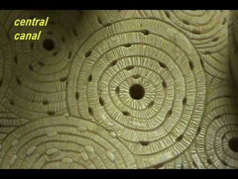
Exploration of the Human Spinal Cord
Amazon.com: My First Lab Duo Scope Microscope - Young … KIDS MICROSCOPE KIT -- This kids microscope kit, in addition to the user manual and experiment guide, includes 5 blank slides, 1 concavity (well) slide, 4 prepared slides, 2 bottles of stain, 5 slide labels, 5 cover glasses, 50 sheets of lens paper, 1 plastic transfer pipette, 1 plain wooden applicator, 1 cotton-tipped applicator, 1 plastic forceps, 1 plastic test tube and 1 plastic …
1.1 Labelling Microscope - Labelled diagram
Label the microscope — Science Learning Hub All microscopes share features in common. In this interactive, you can label the different parts of a microscope. Use this with the Microscope parts activity to help students identify and label the main parts of a microscope and then describe their functions. Drag and drop the text labels onto the microscope diagram.

Stacey Kalkowski's Art Journal: Pollen Spores and Asteroids
ZEISS Axiocam Microscope Cameras for Science and Research For easy documentation, some digital cameras can record images either completely without a computer, or through a connected PC, laptop or iPad running the ZEISS Labscope software. All our microscope cameras are fully supported in our ZEN software environment, offering fast live image display and easy-to-use user interface.

Beyond the Human Eye: A tiny aquatic worm that clones itself
Compound Microscope - Diagram (Parts labelled), Principle and Uses Compound Microscope - Diagram (Parts labelled), Principle and Uses As the name suggests, a compound microscope uses a combination of lenses coupled with an artificial light source to magnify an object at various zoom levels to study the object. A compound microscope: Is used to view samples that are not visible to the naked eye

Amoeba proteus 400x's | Nucleus and pseudopods clear | Flickr - Photo Sharing!
PDF Label parts of the Microscope Label parts of the Microscope: . Created Date: 20150715115425Z
Post a Comment for "43 microscope images with labels"