45 human eye diagram without labels
Label Parts of the Human Eye - University of Dayton Select the correct label for each part of the eye. The image is taken from above the left eye. Click on the Score button to see how you did. Incorrect answers will be marked in red. Human Body Diagram - Bodytomy Human Body Diagram. The human body is one complex network, universally accepted as the most intriguing construct. It is certainly the most widely studied structure the world over. Undermentioned are little- and well-known facts about the human body.
Eye Diagram Unlabelled - Wiring Diagram Pictures Download and use them in your website, document or presentation. Best Human eye diagram unlabelled free vector download for commercial use in ai, eps, cdr, svg vector illustration graphic art design format. human eye. Ask A Biologistcoloring page | Web address:schematron.org coloring. Human Eye. Page 2. 5. 3. 2. 4.

Human eye diagram without labels
Eye Anatomy: 16 Parts of the Eye & Their Functions Apr 05, 2022 · The macula lutea is a yellow oval area in the retina's center (back of the eye). The center of the macula is known as the fovea. It is the section of the retina that is in charge of sharp, detailed central vision (also called visual acuity). The macula lutea has a high concentration of cones. File:Diagram of human eye without labels.svg - Wikimedia Commons Jun 28, 2021 · File:Diagram of human eye without labels.svg. Size of this PNG preview of this SVG file: 410 × 430 pixels. Other resolutions: 229 × 240 pixels | 458 × 480 pixels | 732 × 768 pixels | 976 × 1,024 pixels | 1,953 × 2,048 pixels. Label the Eye Diagram - Enchanted Learning Label the Eye Diagram. Human Anatomy. Read the definitions, then label the eye anatomy diagram below. Cornea - the clear, dome-shaped tissue covering the front of the eye. Iris - the colored part of the eye - it controls the amount of light that enters the eye by changing the size of the pupil. Lens - a crystalline structure located just behind ...
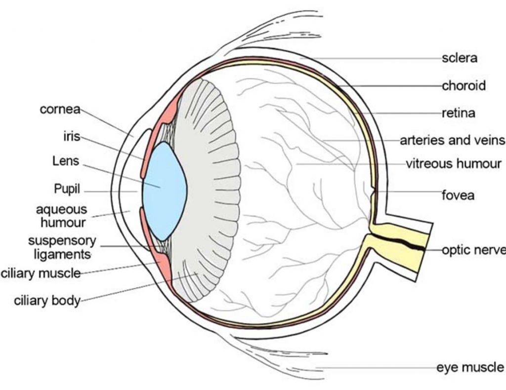
Human eye diagram without labels. Human Body Parts Images Without Labels - Free Vector ... Human ear diagram with labels and label of anatomy labeling the ear purposegames nose diagram with label diagrams all labels human ear the ear diagram without labels anatomy human charts. Illustration Of Body Parts Labels It is certainly the most widely studied structure the world over. Human body parts images without labels. Download body ... Eye Diagram With Labels and detailed description - BYJUS A brief description of the eye along with a well-labelled diagram is given below for reference. Well-Labelled Diagram of Eye The anterior chamber of the eye is the space between the cornea and the iris and is filled with a lubricating fluid, aqueous humour. The vascular layer of the eye, known as the choroid contains the connective tissue. 43 diagram of the human eye without labels 43 diagram of the human eye without labels May 16, 2022 Human Eye - Definition, Structure, Function, Parts, Diagram A human eye is roughly 2.3 cm in diameter and is almost a spherical ball filled with some fluid. Human Ear Diagram - Bodytomy Look no further, this Bodytomy article gives you a labeled human ear diagram and also explains the functions of its different components. The human body is like a big machine, and various processes take place inside it. With the help of the various organs and tissues, it carries out some of the most marvelous tasks, that are no less than a ...
Eye Diagram - Differentiated Worksheets and ... - Pinterest Description Use these simple eye diagrams to help students learn about the human eye. Three differentiated worksheets are included: 1. Write the words using a word bank 2. Cut and paste the words 3. Write the words without a word bank Labels include: eyebrow, eyelid, eyelashes, pupil, iris, and sclera. Eye Test: 3 Free Eye Charts to Download and Print at Home Eye doctors can use different eye test charts for different patients and situations. The three most common eye charts are: Snellen eye chart. "Tumbling E" eye chart. Jaeger eye chart. We've included a link to download your very own eye chart after each section below. You can print these charts and test your vision right in your own home. FREE! - Label the Eye Worksheet - Teacher-Made Learning ... The first page is a labelling exercise with two diagrams of the human eye. One is a view from the outside, and the other is a more detailed cross-section. On the second page, you'll find a set of answers showing the properly labelled human eyes, designed to help you check the worksheets without having to come up with your own answer key. Human eye diagram Images, Stock Photos & Vectors - Shutterstock Human eye diagram royalty-free images 6,729 human eye diagram stock photos, vectors, and illustrations are available royalty-free. See human eye diagram stock video clips Image type Orientation Sort by Popular Biology Healthcare and Medical human eye anatomy eye retina medicine visual perception cone cell pupil Next of 68
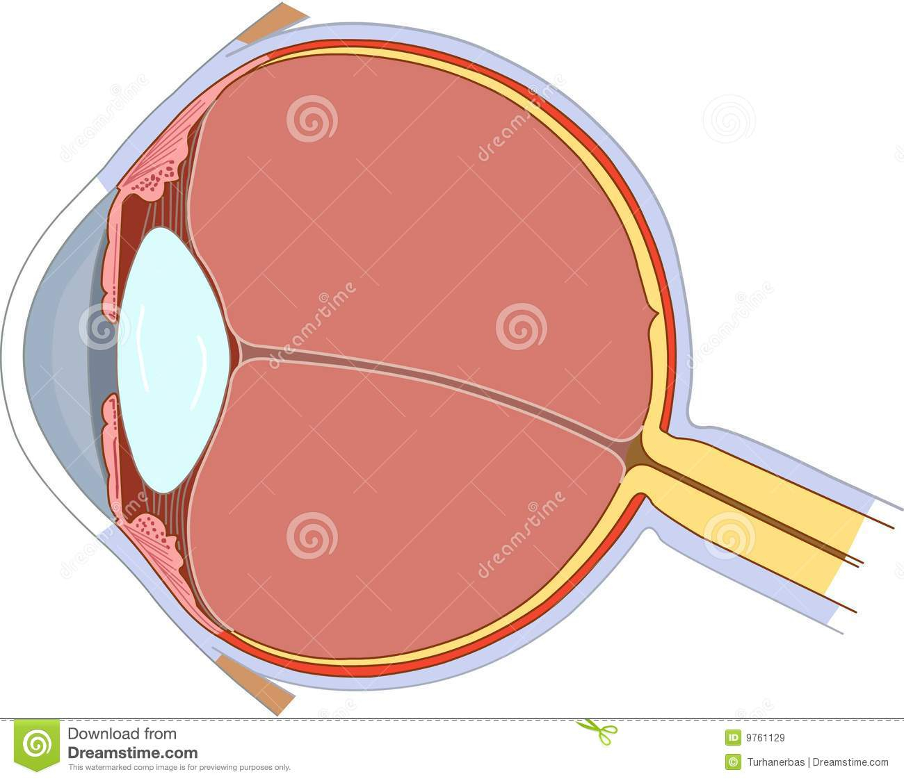
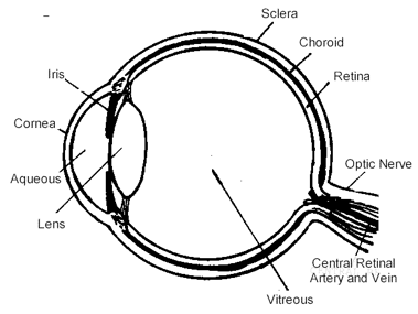
Parts of the Eye - National Eye Institute Eye Diagram Handout Author: National Eye Health Education Program of the National Eye Institute, National Institutes of Health Subject: Handout illustrating parts of the eye Keywords: parts of the eye, eye diagram, vitreous gel, iris, cornea, pupil, lens, optic nerve, macula, retina Created Date: 12/16/2011 12:39:09 PM Human eye - Wikipedia Schematic diagram of the human eye. It shows a horizontal section through the right eye. The eye is made up of three coats, or layers, enclosing various anatomical structures. The outermost layer, known as the fibrous tunic, is composed of the cornea and sclera, which provide shape to the eye and support the deeper structures. Anatomy of the eye: Quizzes and diagrams - Kenhub Oct 28, 2021 · Take a look at the diagram of the eyeball above. Here you can see all of the main structures in this area. Spend some time reviewing the name and location of each one, then try to label the eye yourself - without peeking! - using the eye diagram (blank) below. Unlabeled diagram of the eye The Eyes (Human Anatomy): Diagram, Optic Nerve, Iris ... Articles On Eye Basics. Your eye is a slightly asymmetrical globe, about an inch in diameter. The front part (what you see in the mirror) includes: Iris: the colored part. Cornea: a clear dome ...

Eye Diagram Blank - Human Anatomy
File:Schematic diagram of the human eye no.svg - Wikimedia ... The following 36 pages use this file: File:Diagram of human eye without labels.svg; File:Oeil2.jpg; File:Schematic diagram of anterior segment human eye.svg

Label Parts of the Human Ear
PDF Anatomy of Heart Labeled and Unlabeled Images B Fibrous pericardium Serous pericardium: Parietal pericardium Visceral pericardium (epicardium) Pericardial cavity (b) Frontal dissection of the heart and pericardial cavity
parts of the eyes clipart 20 free Cliparts | Download images on Clipground 2021
Human Eye Diagram, How The Eye Work -15 Amazing Facts of Eye FACT 12 The human eye can detect more than 10 million different types of colors. FACT 13 The world’s most common eye colour is brown. FACT 14 The average person blinks 12 times a minute. Scientists have estimated that we blink between 20,000 and 30,000 times per day on average. FACT 15 Dogs cannot distinguish between GREEN and RED.

Human Eye Diagram To Label Ks2 - Food Ideas
Eyes - Layers of Learning | Human eye diagram, Parts of ... Human Eye Diagram Ear Diagram Science Student Kindergarten Science Science For Kids Science Tools Science Ideas Elementary Science Description Use these simple eye diagrams to help students learn about the human eye. Three differentiated worksheets are included: 1. Write the words using a word bank 2. Cut and paste the words 3.
Schematic Diagram Eye Human Anatomy Labeled Stock Illustration 298561235 - Shutterstock
Eye, External Front View - resource - Imageshare Eye, External Front View. Diagram of the external view of a human eye. Design modalities for the image include braille with and without labels, print with and without labels in greyscale, color, and texture. (Source: Benetech)
Human Eye Anatomy Images, Stock Photos & Vectors | Shutterstock
The Human Eye | Boundless Physics - Lumen Learning The fundus is on the opposite of the pupil, but inside the eye and can not be seen without special instruments. The optic nerve is what conveys the signals of the eye to the brain. is a diagram of the eye. The human eye is made up of three coats: Diagram of the Human Eye: The cornea and lens of an eye act together to form a real image on the ...

File:Schematic diagram of the human eye is.svg - Wikimedia Commons
The Eye Diagram: What is it and why is it used? The eye diagram takes its name from the fact that it has the appearance of a human eye. It is created simply by superimposing successive waveforms to form a composite image. The eye diagram is used primarily to look at digital signals for the purpose of recognizing the effects of distortion and finding its source.

The Eye - Science Quiz
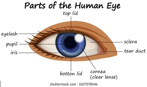
Eye Anatomy: Parts of the Eye and How We See - American ... Here is a tour of the eye starting from the outside, going in through the front and working to the back. Eye Anatomy: Parts of the Eye Outside the Eyeball. The eye sits in a protective bony socket called the orbit. Six extraocular muscles in the orbit are attached to the eye. These muscles move the eye up and down, side to side, and rotate the eye.

Labeled Diagram Of An Eye - Eye Anatomy A Closer Look At The Parts Of The Eye - Just the ...
PDF Eye Anatomy Handout - National Eye Institute of light entering the eye. Lens: The lens is a clear part of the eye behind the iris that helps to focus light, or an image, on the retina. Macula: The macula is the small, sensitive area of the retina that gives central vision. It is located in the center of the retina. Optic nerve: The optic nerve is the largest sensory nerve of the eye.

13 best Eye Diagrams images on Pinterest | Eyes, Eye anatomy and Human anatomy
Human Eye Anatomy - Parts of the Eye ... - All About Vision Eye anatomy: A closer look at the parts of the eye. By Liz Segre. When surveyed about the five senses — sight, hearing, taste, smell and touch — people consistently report that their eyesight is the mode of perception they value (and fear losing) most. Despite this, many people don't have a good understanding of the anatomy of the eye, how ...
Eye Doctors, Lasic Surgery | Chapel Hill, Durham, NC » How the Eye Works
Structure and Functions of Human Eye with labelled Diagram The human eye is a roughly spherical organ, responsible for perceiving visual stimuli. It is enclosed within the eye sockets in the skull and is anchored down by muscles within the sockets. Anatomically, the eye comprises two components fused into one; hence, it does not possess a perfect spherical shape.

picture front of the eye without labels clipart - Clipground
Ear Diagram Unlabeled - wiringall.com Best Unlabeled diagram human ear free vector download for commercial use in ai, eps, cdr, svg vector illustration graphic art design format. unlabeled. Test students' knowledge of the human eye and ear as they color and label these diagrams.
parts of the eyes clipart - Clipground
Label the Eye Diagram - Enchanted Learning Label the Eye Diagram. Human Anatomy. Read the definitions, then label the eye anatomy diagram below. Cornea - the clear, dome-shaped tissue covering the front of the eye. Iris - the colored part of the eye - it controls the amount of light that enters the eye by changing the size of the pupil. Lens - a crystalline structure located just behind ...
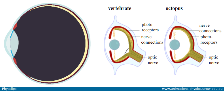
Eye:optics, anatomy and accommodation: Physclips - Light
File:Diagram of human eye without labels.svg - Wikimedia Commons Jun 28, 2021 · File:Diagram of human eye without labels.svg. Size of this PNG preview of this SVG file: 410 × 430 pixels. Other resolutions: 229 × 240 pixels | 458 × 480 pixels | 732 × 768 pixels | 976 × 1,024 pixels | 1,953 × 2,048 pixels.

Schematic Diagram of the Human Eye Assignment
Eye Anatomy: 16 Parts of the Eye & Their Functions Apr 05, 2022 · The macula lutea is a yellow oval area in the retina's center (back of the eye). The center of the macula is known as the fovea. It is the section of the retina that is in charge of sharp, detailed central vision (also called visual acuity). The macula lutea has a high concentration of cones.

How the eye works - Medical Information Illustrated

picture front of the eye without labels clipart - Clipground
Post a Comment for "45 human eye diagram without labels"